Contributed by: Steven J. Rockoff, MD and Diana L. Lam, MD – June 1, 2020
Question 1
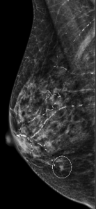
A 56-year-old woman presents for a screening mammogram:


How do you best describe the patient’s breast density?
A. The breasts are almost entirely fatty
B. There are scattered areas of fibroglandular density
C. The breasts are heterogeneously dense
D. The breasts are extremely dense
Answer
C. The breasts are heterogeneously dense
Explanation: One of the first steps in interpreting a mammogram is assessing the breast composition, also known as breast density. There are four acceptable categories described in the ACR BI-RADS manual, which are listed above. This patient’s breasts are heterogeneously dense on mammogram, which may obscure small masses.
Question 2
A representative zoomed in image is shown here:

What is the dominant type of calcification that is present?
A. Fine pleomorphic calcifications
B. Large rod-like calcifications
C. Vascular calcifications
D. Linear calcifications
Answer
C. Vascular calcifications
Explanation: These are benign vascular calcifications, present in a diffuse distribution bilaterally. These have the characteristic “tram-track” appearance.
Note that “linear” is not a type of calcification like the other answer choices, but rather is a descriptor for the distribution of calcifications.
Question 3
Although difficult to appreciate on the 2-D whole breast images provided above, there was a screen-detected mass in the lower inner quadrant of the right breast. Here is the mass noted on MLO and spot tomosynthesis CC images:

What is the best descriptor for the margins of this mass on mammogram?
A. Circumscribed
B. Obscured
C. Microlobulated
D. Indistinct
E. Spiculated
Answer
E. Spiculated
Explanation: The numerous thin lines radiating outward from the mass are indicative of spiculated margins, which is a suspicious finding.
A mass with circumscribed margins has at least 75% of its margins sharply demarcated and separable from the surrounding tissue.
A mass with obscured margins has at least 25% of its margin hidden by superimposed or adjacent breast tissue.
A mass with microlobulated margins has multiple small outward undulations.
A mass with indistinct margins has no clear demarcation of its margin from the surrounding tissue.
Question 4
Targeted ultrasound was performed, with a representative image of the mass shown here:
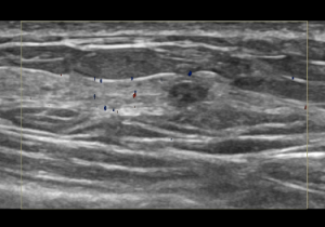
What is the best descriptor for the margins of this mass on ultrasound?
A. Indistinct
B. Angular
C. Microlobulated
D. Spiculated
Answer
A. Indistinct
Explanation: This mass is not circumscribed on ultrasound. The margin between the majority of the mass and surrounding tissue is not clearly defined, and there is a lack of other features that would be seen with the other margin descriptors. This mass’s sonographic margin is best described as indistinct.
Ultrasound-guided biopsy was performed, which yielded invasive ductal carcinoma.