Contributed by: Steven J. Rockoff, MD and Diana L. Lam, MD – June 1, 2020
Question 1
A 48-year-old woman presents for screening mammogram:
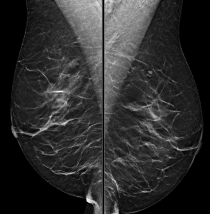

How do you best describe the patient’s breast density?
A. The breasts are almost entirely fatty
B. There are scattered areas of fibroglandular density
C. The breasts are heterogeneously dense
D. The breasts are extremely dense
Answer
Either B. There are scattered areas of fibroglandular density, or C. The breasts are heterogeneously dense
Explanation: One of the first steps in interpreting a mammogram is assessing the breast composition, also known as breast density. There are four acceptable categories described in the BI-RADS manual, which are listed above. Breast density is a subjective assessment of how much fibroglandular tissue compared to fat tissue is present in the breast, and also has a correlation to how difficult it may be to interpret a patient’s mammogram. Woman who have “dense” breasts (heterogeneously or extremely dense) have a slightly increased risk of developing breast cancer compared to women with non-dense breasts. This patient’s breasts were reported as being composed of scattered areas of fibroglandular density. Since this is a subjective determination, describing them as heterogeneously dense could also be acceptable.
Question 2
A mass is present in the upper outer quadrant of the left breast:

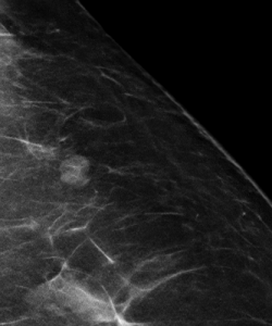
How is this mass best described?
A. BI-RADS 0 (Incomplete); Recommend diagnostic mammogram and ultrasound
B. BI-RADS 2 (Benign); Recommend one year follow-up
C. BI-RADS 3 (Probably Benign); Recommend six month follow-up
D. BI-RADS 4 (Suspicious); Recommend biopsy
Answer
B. BI-RADS 2 (Benign); Recommend one year follow-up
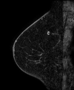
Explanation: This is the classic appearance of an intramammary lymph node on mammography. This is a normal breast structure, typically appearing as a circumscribed small mass on the mammogram with a lucent notch that corresponds to the anatomic fatty hilum. The most common location is in the upper outer quadrant. If the appearance is not quite classic, a history of stability on prior mammograms is reassuring that this is most likely a normal/benign lymph node.
In this patient’s case, her intramammary lymph node had been unchanged for many years and has the expected appearance of a lymph node on this post-contrast MRI image: