Contributed by: Steven J. Rockoff, MD and Diana L. Lam, MD – June 1, 2020
Question 1
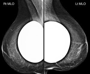
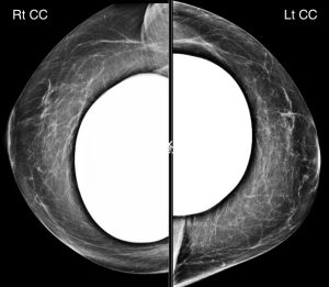
A 50-year-old woman presents for a screening mammogram:
What kind of implants are present?
A. Saline implants
B. Silicone implants
C. Ceramic implants
Answer
B. Silicone implants
Explanation: The extremely high and homogenous density of these implants indicates that they are composed of silicone.
With saline implants, we should be able to see “through” and identify the implant valves.
Question 2
There is a unilateral finding present. What is it?
A. Right breast retroareolar mass
B. Right breast extracapsular implant rupture
C. Left breast lower outer quadrant fat-containing mass
D. Left axillary mass
Answer
C. Left breast lower outer quadrant fat-containing mass
Explanation: The fat-containing mass shown below is present in the lower outer quadrant of the left breast.
Mammography is not a sensitive modality to assess for silicone implant rupture, although there is no gross evidence of extracapsular silicone implant rupture on these images. Saline implant rupture is a clinical diagnosis.
Question 3

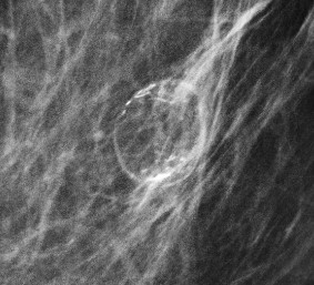
What is the best descriptor for the type of calcification that is seen with this fat-containing mass?
A. Rim calcification
B. Round calcification
C. Fine linear branching calcification
D. Popcorn-like calcification
Answer
A. Rim calcification
Explanation: A rim calcification is composed of a continuous rim of thin (≤ 1 mm) calcification that surrounds a round or oval structure, usually with distinct internal fat density, as is true for this case. This large rim calcification has the typical appearance of fat necrosis, as can be seen in the setting of prior breast trauma or surgery (such as implant placement). Oil cysts or simple cysts (not shown in this case) can also have smaller rim calcifications. Rim calcifications are typically benign.