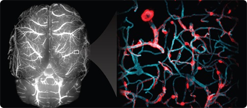
The Shih Lab uses advanced optical imaging to study neurovascular function in the living brain. Our goal is to better understand how blood flows through the brain by watching and learning from model organisms. This can provide clues on the development and repair of key vascular functions, such as the blood-brain barrier, which may one day help protect blood vessels in the aging and diseased brain.
Our research currently has two main focuses:
- We are interested in how cerebral microinfarcts contribute to cognitive impairment and dementia. Microinfarcts are small but surprisingly common lesions in the aging brain. They are thought to arise from the clogging of tiny blood vessels during small vessel disease. Despite their small size (about the size of a pinhead), microinfarcts can number in the hundreds to thousands. They can affect the brain in regions beyond where the injury actually occurs, contributing broadly to brain dysfunction. To better understand microinfarcts, we model them in the rodent brain by optically occluding small brain vessels. We then use a combination of in vivo two-photon imaging, high-field MRI and histology to quantify their effects on neuronal connectivity, inflammatory responses and neurovascular function. We also test potential therapeutics that are able to mitigate microinfarct injury.
- We are also interested in the role of brain pericytes in cerebrovascular function. Pericytes, along with endothelial cells, are an integral component of the vascular wall. During brain development, pericytes help to construct the blood-brain barrier, and build intricate vascular networks for delivering blood to all brain cells. During aging and especially in Alzheimer’s disease, there is an accelerated loss of pericytes that may contribute to exacerbation of the disease. Our lab is using in vivo two-photon imaging to study this fascinating cell type in comparative studies of pericytes in the young, developing brain versus the aging brain.