Imaging
Hardie, MacDonald and Rubel, Brain Research Methods
6015s3
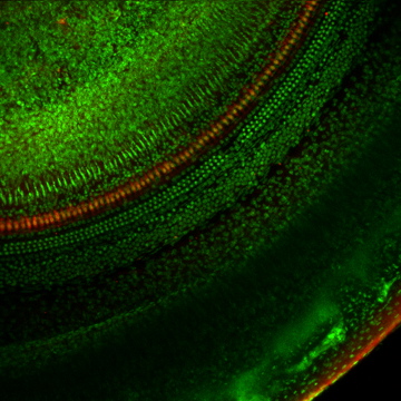
LSCM image stack collected from the organ of Corti, C57BL6 mouse, 2
months old, imaged by combined autofluorescence and the DNA label, Yo-Pro-1.
Nuclei are brightly visible against paraformaldehyde induced tissue
autofluorescence. Note that the well formed anatomy allows ready identification
of the cell types and 9 IHC nuclei. Compare this to movies from animal
#6421. 488 nm Excitation/522DF35 Emission filter. LSCM, Nikon 60X PlanApo,
Zoom=2, 163 slices, 0.4 µm per slice.
6015cmuls3
Brightest point projections of the same image stack as in 6015s3, using
the Object Image “Fire” look-up table. Nuclei are brightly
colored against the blue autofluorescence. A line has been drawn along
the row of IHC nuclei to measure the cochlear duct. 488 nm Excitation/522DF35
Emission filter. 65.2 µm thick optical volume.
64216201
LSCM image stack of combined autofluorescence and Yo-Pro-1 nuclear label
from a 15 month old C57BL6 mouse. Note that the distorted anatomy of
the collapsed organ of Corti makes it impossible to determine presence
of IHC nuclei below the tectorial membrane. 488 nm Excitation/522DF35
Emission filter. LSCM, Nikon 60X PlanApo, Zoom=2, 152 slices, 0.4 µm
per slice.
6421cmuls3
Brightest point projections of the same image stack as in 6421s3, using
the Object Image “Fire” look-up table. Nuclei are brightly
colored against the blue autofluorescence. A line has been drawn along
the basilar lamina below the approximate position of the IHC.. 488 nm
Excitation/522DF35 Emission filter. 60.8 µm thick optical volume.
6015VBs3
Surface rendering of the 6015s3 stack, with the autofluorescence emissions
collected in 2 channels with 522DF35 and 605DF32 filters. Pixel intensities
from autofluorescence are in the green channel and pixels from nuclear
label are in the red channel. Rendering was carried out with Voxblast.
6421VBs3
Surface rendering of the 6421s3 stack, with the autofluorescence emissions
collected in 2 channels with 522DF35 and 605DF32 filters. Pixel intensities
from autofluorescence are in the green channel and pixels from nuclear
label are in the red channel. Rendering was carried out with Voxblast.
36410lsms3
Modiolar view from a C57BL6 mouse, 21 days old, acquired by multi-photon
LSCM. IHC somata in the upper turn are labeled by calretinin (red).
Nerve fibers innervating the IHCs and OHCs are labeled for neurofilaments
(green). Multi-photon imaging appears to have less signal attenuation
through the depth of the volume. Zeiss 40X PlanNeoFluor, Zoom = 2, 38
µm optical volume.
36416T2Prestins3b
Outer hair cells are labeled by antibodies to prestin (green), IHCs
are labeled for calretinin (red) while To-Pro-3 identifies nuclei (blue)
in the organ of Corti from a 21 day old C57BL6 mouse. LSCM, Nikon 60X
PlanApo, Zoom = 2, 28.8 µm optical thickness.
303T01calrets3
Nerve fibers and IHCs are labeled by calretinin in a surface view of
the lower turn from a 21 day old C57BL6 mouse. Nerves constrict as they
pass through the habenula perforata then spread to innervate the IHCs.
LSCM, Nikon 60X PlanApo, Zoom = 1, optical volume is 82.5 µm thick.
6431T2s3b
Modiolar view of the organ of Corti from an aged C57BL6 mouse displays
missing IHCs in the lower turn. IHC somata and nerves are labeled for
calretinin (red) while beta-tubulin (green) labels support cells, oligodendrocytes,
basal lamina and Hensen’s cells. Thin crossing fibers may be observed
in the tunnel of Corti. LSCM, Nikon 60X PlanApo, Zoom = 2, 60 µm
thick optical volume, 78.2 µm cochlear duct length.
60164ys3
The gross anatomy and spiral of the cochlear duct from a C57BL6 mouse,
aged 60 days, acquired by LSCM, are presented in a combined image of
autofluorescence and Yo-Pro-1 nuclear label. Nikon 4X EPlan, Zoom=1,
optical volume of 200 µm thickness.