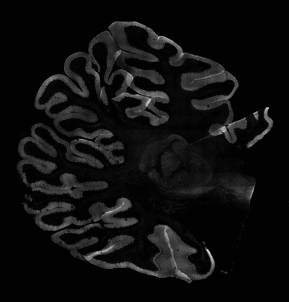
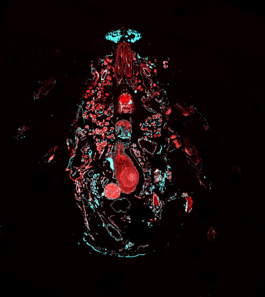
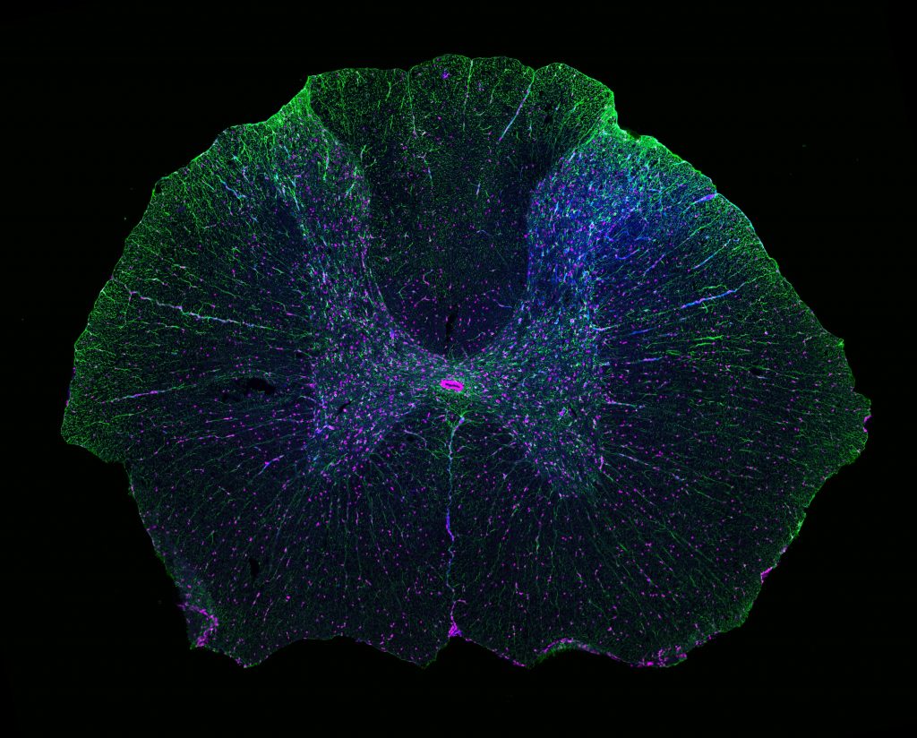
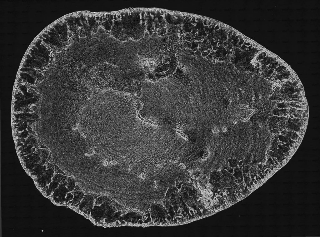
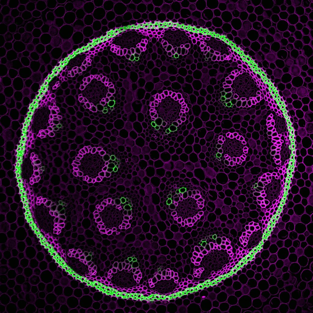
The following images were acquired on Keck Center microscopes by the Keck Center Manager, Dr. Nathaniel Peters, working alongside Keck Center users from various UW research labs. Links to lab websites can be found in the image captions. Images may not be reproduced or reused without permission.

Purkinje Cells in a section of Rhesus macaque Cerebellum – Horwitz Lab (UW) – Confocal Mosaic Tilescan
Cross-Section of a Tick – Mougous Lab (UW) – Confocal Mosaic Tilescan
3 Stages of Drosophila melanogaster Egg Chamber Development, E-Cadherin::GFP – Berg Lab (UW) – Maximum Intensity Projections of Confocal Z-Series at 3 Time Points
Rattus norvegicus Spinal Cord Section – Hofstetter Lab (UW) – Confocal Mosaic Tilescan
A Stage 12 Drosophila melanogaster Egg Chamber Stained for DNA (yellow) and Dynamin Protein (red) – Berg Lab (UW) – Maximum Intensity Projections of a Confocal Z-series
Air Pockets in Solidified Mounting Medium Surrounding a Piece of Debris – Darkfield Mosaic Tiling
A Stage 12 Drosophila melanogaster Egg Chamber Stained for DNA and paxillin mRNA – Berg Lab (UW) – Maximum Intensity Projections of a Confocal Z-series
A Dried Droplet of a Water-Phospholipid Suspension – De Yoreo and Keller Labs – Phase Contrast Mosaic Tiling

Convallaria majalis (Lily of the Valley) Root Tip Cross Section – Confocal Mosaic Tilescan
Drosophila melanogaster Egg Chamber Stained for DNA (blue), Broad Protein (cyan), Rhomboid-lacZ (red), and E-Cadherin (magenta) – Berg Lab (UW) – Maximum Intensity Projections of a Confocal Z-series