Widefield Microscopes
The Keck Center’s widefield microscope systems use fluorescent lamp excitation, filter cubes, and CCD monochrome and/or RGB-color cameras to produce fluorescent, brightfield, DIC, Phase Contrast, and/or RGB-color images. The motorized XY-stage on the Leica DMI6000 microscope allows for mosaic tiling of large tissue sections, both fluorescent and color-stained, and multi-point live imaging. Widefield microscopes do not exclude out-of-focus light as scanning confocal microscopes do, so they are usually not suitable for imaging thick samples.
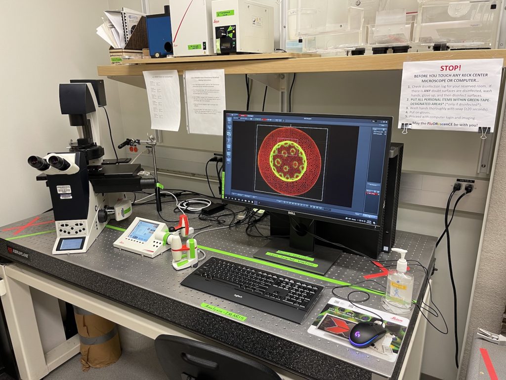
Leica Inverted Multi-Dimensional Widefield
- Leica DMI6000 inverted microscope
- 5X LWD, 10X LWD, 20X, 20X LWD and 40X LWD dry objectives
- 40X and 100X Oil objectives
- 63X water objective
- BFP (blue), CFP (cyan), GFP (green), YFP (yellow), TexasRed (red), and CY5 (far red) filter cubes
- Motorized filters, shutters, and Z-drive
- Motorized stage for mosaic tiling and/or multi-point live-imaging
- DIC optics
- Phase Contrast optics (for use with long working distance (LWD) objectives)
- Z-sectioning
- Mosaic tiling
- Multi-point acquisition for live imaging
- Leica DFC365 FX CCD camera (fluorescence, brightfield, DIC, phase contrast)
- Leica MC170HD Color camera (color – e.g., crystal violet, histological stains)
- LASX software with Navigator
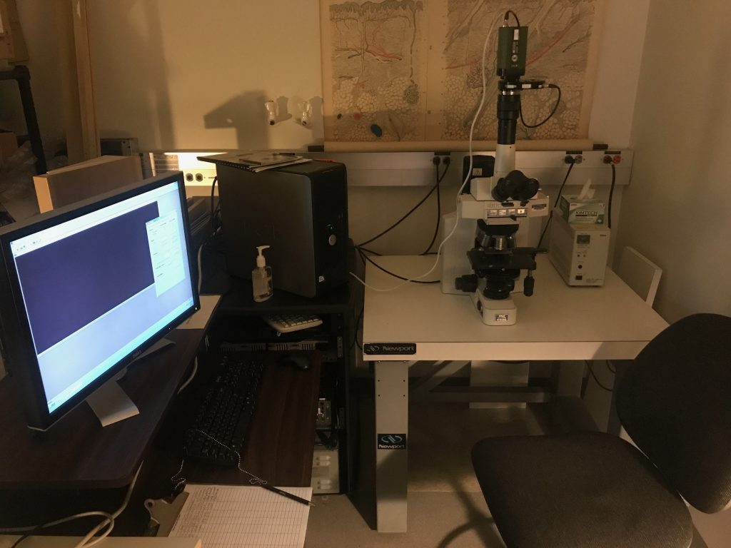
Nikon Upright Widefield
(currently a backup system, inquire for access)
- Nikon Eclipse E600 upright microscope
- Manual Stage
- 1X, 2X, 4X, 10X, 20X, and 40X dry objectives
- 63X Oil objective
- Blue, green, and red filter cubes
- DIC optics
- Q-Imaging Retigia EX CCD Camera (monochrome) with optional color filter for imaging histology slides
- Q-Capture software