
Pathology in a Tube for 3D optical imaging

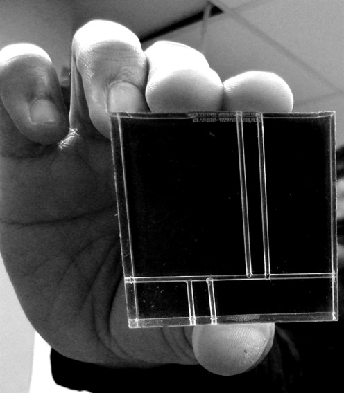
Prototype milli-fluidic device for staining a fresh core needle biopsy.
Das, R., Kramer, G.M., Burfeind, C.W., and Seibel, E.J. (2014) Pathology in a tube, step 1: fixing, staining, and transporting pancreatic core biopsies in a microfluidic device for 3D imaging, Microfluidics, BioMEMS, and Medical Microsystems XII, Proc. SPIE vol. 8976, paper 89760R-1-8.
Das, R., Burfeind, CW, Lim, SD, Patle, S., Seibel, EJ. (2018) Pathology in a tube: Step 2. Simple, rapid fabrication of curved, circular cross section millifluidic channels for biopsy preparation/3D imaging towards pancreatic cancer diagnosis, Proc. SPIE 10491, Microfluidics, BioMEMS, and Medical Microsystems XVI, 1049118 (19 February 2018); doi:10.1117/12.2291018.
CoreView – Core Needle Biopsy Assessment
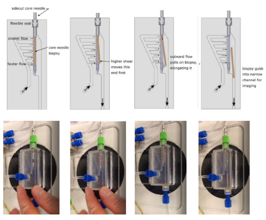
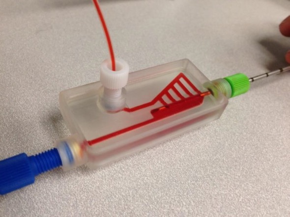
needle inserted and red dye added for visualization.
Cooper, DJ, Fauver, ME, Dintzis, SM, and Seibel, EJ. (2020) Rapid Needle Biopsy Assessment at Point of Care to Advance Personalized Cancer Therapy, AACR Annual Meeting 2020, virtual presentation, June virtual presentation.