Advanced Physical Diagnosis
Examination
  • Techniques
• 1st & 2nd Heart Sounds
• 2nd & 3rd Heart Sounds
• Clicks and Snaps
• Murmurs
• Rubs
  • Demonstrations
Historical
Pathophysiology
Associated Evaluations
  • Patient HX
  • Physical Exam
  • Laboratory & Imaging
Differential Dx
Evidence Base
• Accuracy in Diagnosis of Systolic Murmurs
• Accuracy in Diagnosis of Diastolic Murmurs
• Accuracy in Diagnosis of CHF
References
Teaching Tips
[Skill Modules >> Heart Sounds & Murmurs >> Techniques ]

Techniques: Heart Sounds & Murmurs

Third & Fourth Heart Sounds

Click to hear S3 extra heart sound. Click to hear S4 extra heart sound.


Diagram of S3 S4 heart sounds during systole and diastole.

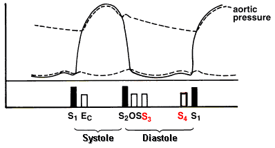
A triple rhythm in diastole is called a gallop and results from the presence of a S3, S4 or both.

Description:
Both sounds are low frequency and thus best heard with the bell of the stethoscope.

Location:
If originating from LV

If originating from RV

  • Usually best heard over left lower sternal border
  • Louder during inspiration

back to top

Third Heart Sound S3

Description:
Low frequency sound in early diastole, 120 to 180 ms after S2

Sounds like:
Lub du bub S1S3S2 cadence similar to "Kentucky"

Clinical Significance:
Results from increased atrial pressure leading to increased flow rates, as seen in congestive heart failure, which is the most common cause of a S3. Associated dilated cardiomyopathy with dilated ventricles also contribute to the sound. See Accuracy in Diagnosis of CHF .

Less commonly, valvular regurgitation and left to right shunts may also result in a S3 due to increased flow.

May be normal physiological finding in patients less than age 40.

back to top

Fourth Heart Sound S4

Description:
Low frequency sound in presystolic portion of diastole,

Sounds like:
Belub dup S1S4S2 cadence similar to "Tennessee"


Clinical significance:
Seen in patients with stiffened left ventricles, resulting from conditions such as hypertension, aortic stenosis, ischemic or hypertrophic cardiomyopathy.

In patient with mitral regurgitation, suggestive of acute onset of regurgitation due to the rupture of the chorda tendinae that anchor the Valvular leaflets.

Proceed to the next technique

back to top