|
Research Feature Stories: Other News: |
Researchers Able to See Brain in Action
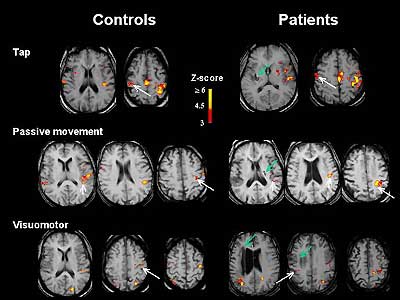
Dr. Steven Cramer, assistant professor of neurology, uses functional magnetic imaging to learn how the brain can restructure itself in response to experience and disease. In this case, images of the living brain anatomy during a simple physical activity—such as when a stroke patient taps a finger—are shedding light on how the brain adapts after a stroke. These functional imaging studies have shown several different ways the brains of stroke victims compensate for injury.
 |
|
Functional magnetic resonance imaging reveals how parts of the brain activate when a subject performs certain tasks. |
Another investigator, Dr. Stephen Dager, professor of psychiatry and behavioral sciences and of radiology and adjunct professor of bioengineering, heads an autism neuroimaging study. This research is attempting to find evidence of brain development abnormalities associated with autism. Combining several imaging techniques for brain structure and chemistry, researchers are trying to determine what goes awry in the brain development of young children with autism. Dager adds that, in conjunction with other currently funded NIH research projects, neuroimaging of brain chemistry holds promise for understanding and treating other conditions like panic disorder, bipolar affective disorder, and obsessive/compulsive disorder.
![]()
|
| UW AMC Medical Center | UW School of Medicine | Harborview | UW MC | Search UW AMC | UW Home | Contact Us | ©2001-2002, University of Washington Academic Medical Center. All rights reserved. Please honor our copyrights. |
|