By Genevieve Wanucha
As published in Dimensions Magazine - Fall 2019
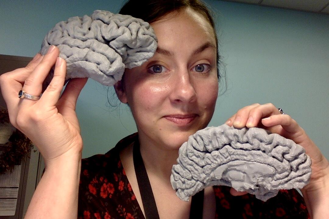
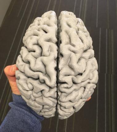
Have you ever wanted to hold your brain in your hands? Well, you can hold mine. It’s a 3D model – at 100% scale and made of a super hard, glimmering grey resin. Its surface is a labyrinth of plump, curving folds. The ‘gyri’ (ridges) and the ‘sulci’ (crevices) rise and fall in convoluted patterns that are different across the two hemispheres, as if to mirror the extroverted introversion of its owner’s personality. The asymmetry reminds me of how abstract, creative mental processes integrate with analytical, logical ones to produce our conscious perception and behavior.
I have wanted a 3D print of my brain ever since I realized it was possible, when research scientist Dan Peterson at the UW Integrated Brain Imaging Center (IBIC) provided several 3D printed brains of Alzheimer’s patients as educational material for UW’s annual Brain Awareness Day, offered to students in grades 2-12. But it wasn’t until now that I had what the IBIC researchers needed to construct my brain model. This May, I got my hands on my magnetic resonance imaging (MRI) scan collected as part of a research study that I participate in each year – and in the specific file format to make it perfectly 3D printable.
Tim Wilbur, research scientist at IBIC, made my brain on his own 3D printer. The printer works by gradually layering segments of the object from the bottom up in thin threads of resin, based on the structural MRI data. The 100% scale brain print took the machine about 70 hours of continuous printing. Before now, our center was limited to brain models printed at just 50% and 75% scale.
“As an MRI technologist I get to see brains of many people, but only in the form of two-dimensional black-and-white pictures,” says Wilbur. “Being able to hold a 3D print of a brain in my hands makes the brain real in a way that the pictures do not. The 3D print also makes it easier to appreciate not only the intricate shapes and contours of the brain structure but also simply the size of the thing, which somehow manages to seem both large – compared to the skull that holds it – and small – considering what it does and what it’s capable of.”
My 3D printed brain, as
Printing 3D brains is a personal project for Wilbur, though it aligns well with his MRI duties at work. He keeps a few different prints of his brain on display in the lab and they serve as talking points for curious research subjects and staff. He recently made a brain print for Dr. Todd Richards, PhD, Emeritus Professor of UW Radiology as a gift in honor of his retirement after 34 years of brain imaging research.
At first, I could not take my eyes off of this model, for I know with certainly that it offers a map, perhaps an unreadable one, of my unique traits, weaknesses, and artistic and creative strengths. Research in behavioral neurology, such as the work of Dr. Bruce Miller, MD, Professor of Neurology at University of California San Francisco, shows that the non-dominant hemisphere (the right hemisphere is non-dominant for the majority of right-handed people) governs the production of visual art. I immediately leverage my position as the science writer for the ADRC, to embark on my most personal scientific journey to date: to decipher the map of my brain folds on that hemisphere. Can I find the basis, the origin of my identity as a creative artist?
An ADRC scientist who looked at my brain model pointed me towards a 2006 publication by neurologists at Beth Israel Deaconess Medical Center and Harvard Medical School, who studied the differences in brain structure in the motor cortex between 16 expert piano players, 16 expert string players, and 32 non-musicians. They found that they could detect anatomical differences by simple visual inspection of 3D renderings of MRI scans.
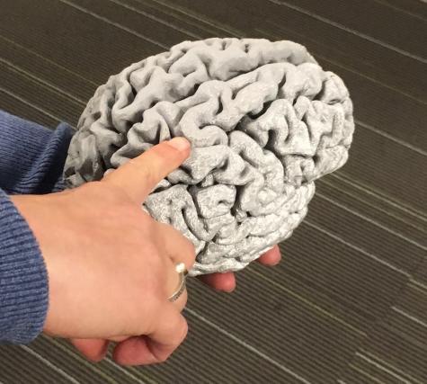
The researchers looked at the precentral gyrus, part of which controls hand and finger movement. The right side of it controls the left hand; the left side of it controls the right hand. This knob of brain tissue, in most people, folds into the characteristic U-shape of an ‘Omega Sign’ on one or both sides of the brain. Studies have found that this brain area is longer and contains denser gray matter in expert musicians than in non-musicians.
When the researchers only looked at the “exceptionally prominent” Omega Signs, they found that the feature appeared more often on the right side in string players and on the left side in piano players. The hypothesis is that years and years of complex left-handed finger movements involved in playing a string instrument thickens up that bit of the right brain, and vice versa for piano playing. (Indeed, neuroanatomical studies of Albert Einstein’s preserved brain, which shows an Omega Sign, attribute this feature to the fact that he took violin lessons as a child.)
A thorough inspection of my 3D brain’s terrain shows I too have an Omega Sign in the right hemisphere (just like 34% of the non-musicians in the study, and 55% of the string players). In case you are wondering, I have never played a musical instrument. If only I had picked up a violin, would I have been a star orchestra player today? Or, more realistically, perhaps the Omega Sign in my right hemisphere has something more fundamental to do with creative expressions, like my art and writing, which require well-controlled and finely detailed hand and finger movements.
Dr. C. Dirk Keene, Associate Professor of UW Pathology who leads the Neuropathology lab at the ADRC, quickly tamped down my self-involved wonderings when I brought the 3D print to his office. “You can think of the folding pattern of a brain like a fingerprint,” he says. “The 3D print is definitely showing you something real. This is not 21st century palm reading, but it’s not really possible to tell, just by looking at it, anything about what makes a person themselves.” (I still think that Omega brain fold is a conduit for my visual creativity.) He told me my brain looked normal.
This is the image caption.
I know that it is not the brain’s structure, but rather a person’s past experiences and resources, and current social and environmental milieu, that influence and ultimately mediate their potential. Still, the human brain creates that tangible sense of ‘me’ and my stable personality – one that may change and transform over time – and that’s incredible. While I reluctantly try to accept that my brain print will never serve as an accurate map of my talents and untapped potential, I am much more certain that the UW ADRC has a very interesting new, thought-provoking tool.
3D brain models, while not a scientific tool, are incredibly valuable in education and communication about neuroscience. “It’s kind of a solution in search of a problem,” says Dan Peterson of IBIC. “One potential idea would be to use the 3D prints as a communication tool during family conferences at the UW Memory and Brain Wellness Center clinic. Another potential application would be to use 3D prints as a ‘thank you’ for participating in research – to build a system where every subject gets a print mailed to them after-the-fact.”
As part of my job at the UW ADRC, I help with community outreach activities and I’m always in need of tools to engage with the audience, often people who have different backgrounds and experiences than me. Even in the short time that I’ve had this model as a tool to bring to events, I‘ve learned that the ability to show my own brain, instead of an anonymous person, has made all the difference in the potential to connect with a diverse array of people about brain science and medicine.
At a recent Alzheimer’s outreach event at the Kalispel Tribe’s Camas Clinic, clinicians and community members at our table took an interest in the brain model, as well as in the ADRC’s slides of brain tissue. Their eyes widened upon realizing they were holding the brain of the person talking to them! At one point, the building security guard came over. I asked him if he wanted to hold the 3D printed brain. “Oh, no I don’t need to do that,” he said. “It’s my brain,” I responded. “…..Well then,” he said, and took the brain into his hands. “Well, isn't this something. How did you manage that?”
A 3D brain is a powerful conversation starter, one that dissolves boredom and naturally provokes emotional responses, surprise, wonder, and reflection, for everyone. I’m looking forward to the next UW Brain Awareness Day in March, where the children always ask us whose brain served as the model for the 3D print. It will be fun to tell them that the owner is standing right in front of them.
For me, this model is much more than an outreach tool, and I’m grateful for the personal journey into neuroanatomy that it sparked. It’s a powerful and comforting experience to hold this object that feels so solid and indestructible; I know how fragile the real thing actually is. As someone who lost loved ones too early to a neurodegenerative disease, holding this beautiful object in my hands creates motivation to use my brain for everything it’s worth, try and take care of it, and make sure I someday figure out where all those curvy paths lead.
__media-listing.png)


